
Michael Hendzel
Principal Investigator
Nuclear BiologyResearch Overview
Internationally recognized leader in cell nucleus, chromatin structure, and genome stability research. Helped demonstrate that condensed chromatin exhibits solid-like behavior at the mesoscale in vitro and in living cells.
The work of the Michael Hendzel lab has reshaped the scientific understanding of genome biology. The lab's 2020 Cell study showed that condensed chromatin exhibits solid-like, elastic gel behavior at the mesoscale in vitro and in living cells, providing a scaffold for liquid-like chromatin-binding proteins. Another major impact came from the lab's 2015 Nature Cell Biology paper identifying the critical role of the RNF138 enzyme in DNA repair.
Research Focus
Genome Stability Mechanisms
The team investigates the complex safeguards that ensure the faithful inheritance of genetic material during cell division. Using advanced single-cell imaging techniques, they observe how cells respond to and cope with DNA damage in real time.
DNA Double-Strand Break Repair
The lab is particularly interested in how a cell chooses between different pathways to repair these highly toxic lesions. They made a breakthrough discovery by identifying the enzyme RNF138 as a key factor that removes a barrier protein from broken DNA ends, thereby promoting the most accurate form of repair.
Chromatin Dynamics & Epigenetic Regulation
This research explores how the physical and chemical state of chromatin affects gene activity and DNA repair. Their findings on the solid-like behavior of condensed chromatin at the mesoscale inform how molecules move within the nucleus and access the genome.
Nuclear Organization
Investigation of how the cell nucleus is organized and how this organization affects DNA repair, gene expression, and cellular function. This work aims to lay the foundation for epigenetic therapies that could make cancer cells more vulnerable to treatment.
Key Publications
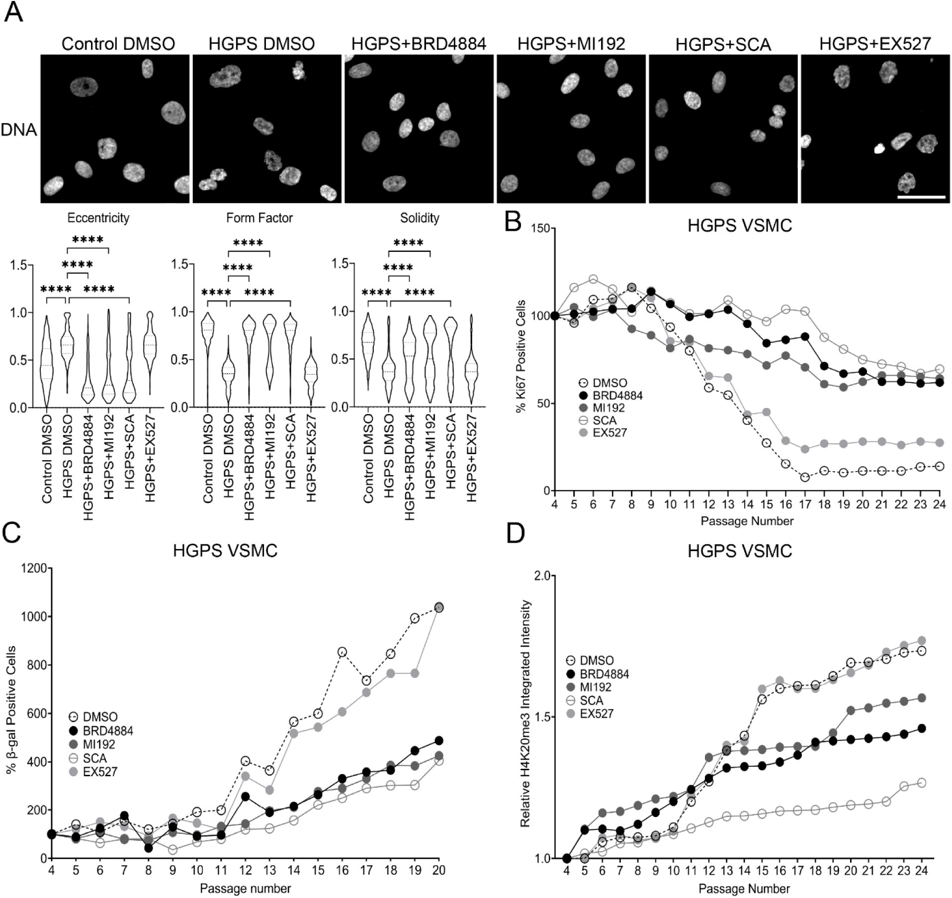
HDAC2 inhibition restores H4K16ac and delays senescence in HGPS VSMCs
Karimpour et al.
This bioRxiv preprint reports cell-culture experiments in control and Hutchinson-Gilford progeria syndrome (HGPS) vascular smooth muscle cells. HDAC2-directed inhibitors restored H4K16 acetylation and improved several cellular senescence-related measures. These findings identify a candidate target for further study; they do not establish clinical efficacy in patients. Read the preprint.
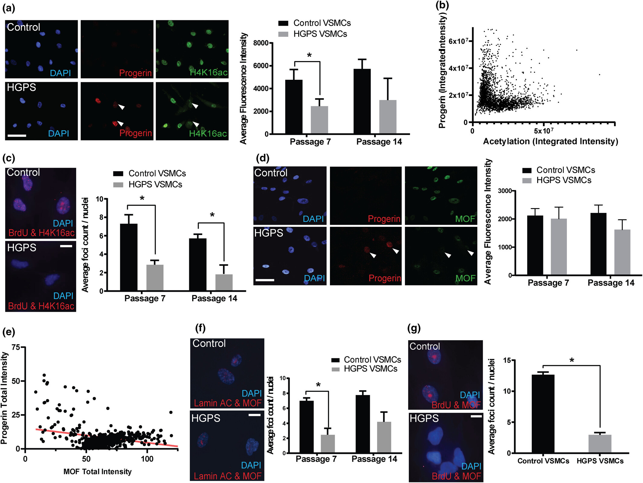
Mechanisms of Vascular Aging in Progeria: Loss of Epigenetic Homeostasis in Vascular Smooth Muscle Cells
Ngubo et al.
Cardiovascular disease is the leading cause of death in HGPS, yet the molecular mechanisms underlying vascular pathology have remained poorly understood. Using patient-derived cells and sophisticated imaging techniques including super-resolution microscopy, we discovered that progerin expression leads to loss of key heterochromatin marks.
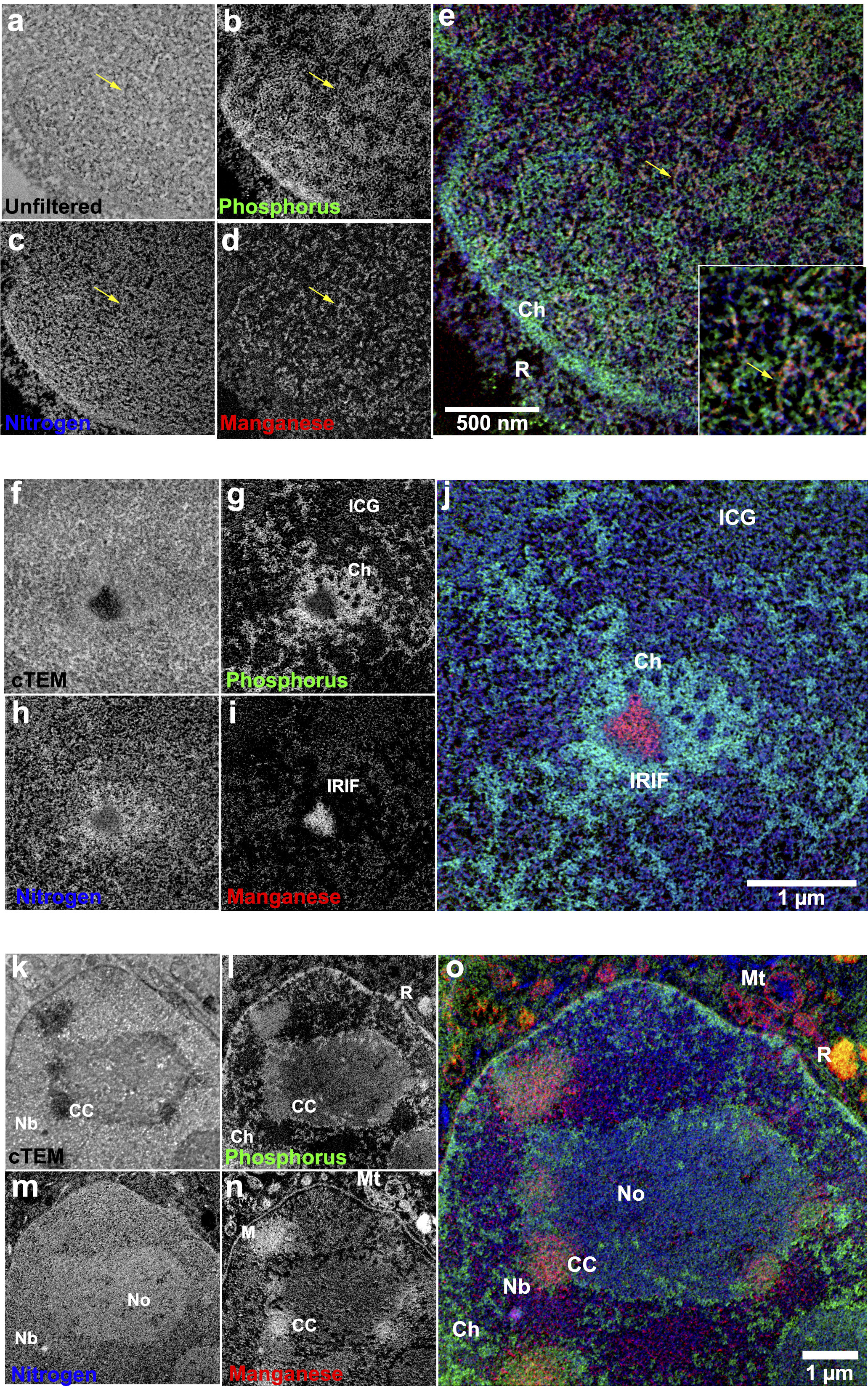
Multichannel Correlative Imaging Reveals Nanoscale Organization of Nuclear Compartments
Booth et al.
This technical tour de force presents a novel correlative imaging approach that combines super-resolution fluorescence microscopy with multi-color electron microscopy to achieve unprecedented visualization of nuclear architecture.
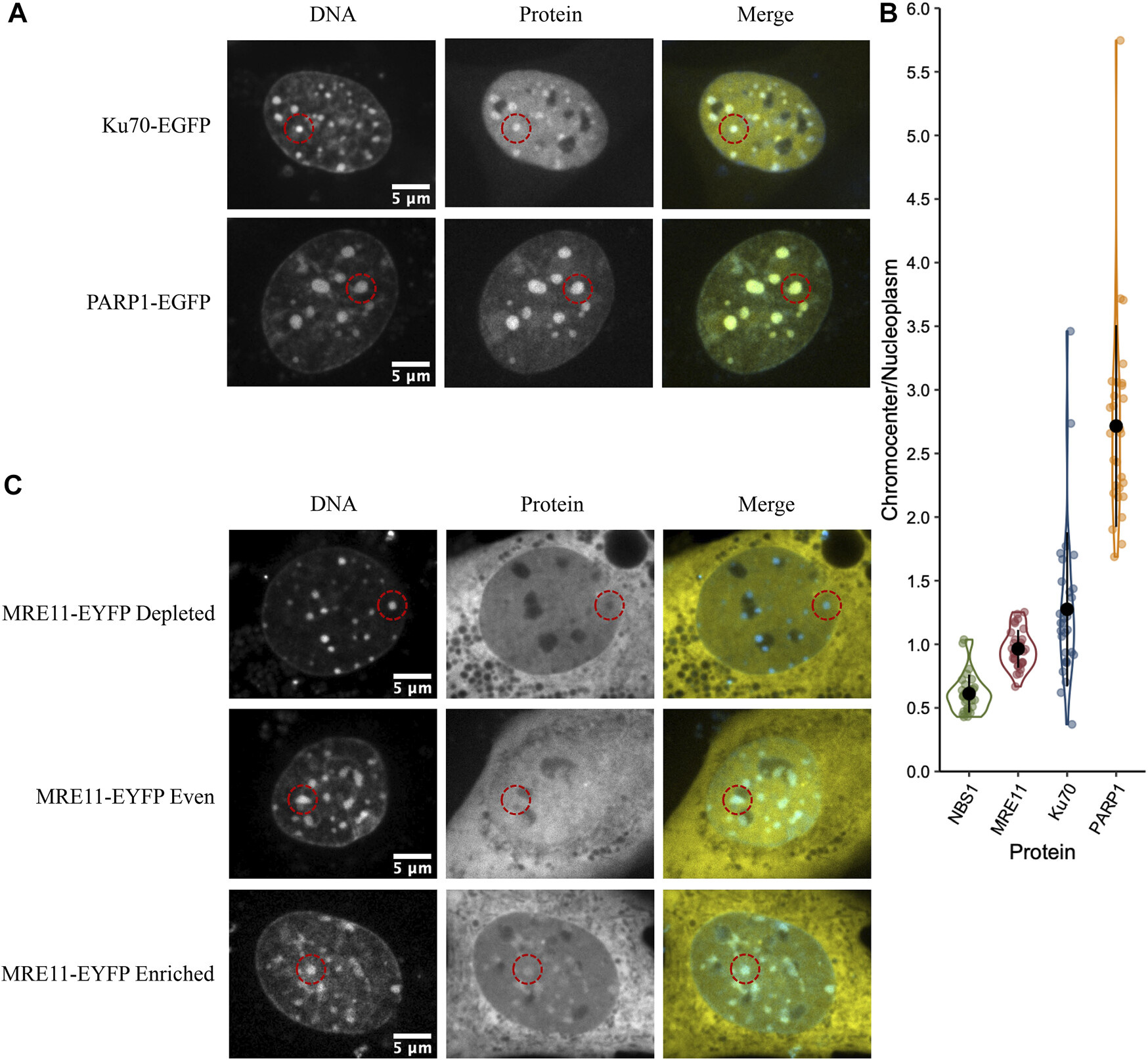
Sub-compartmentalization of DNA Repair Foci
Abate & Hendzel
Using advanced super-resolution microscopy and quantitative image analysis, we discovered that repair foci are not homogeneous structures but contain distinct sub-compartments where different repair proteins are spatially segregated.
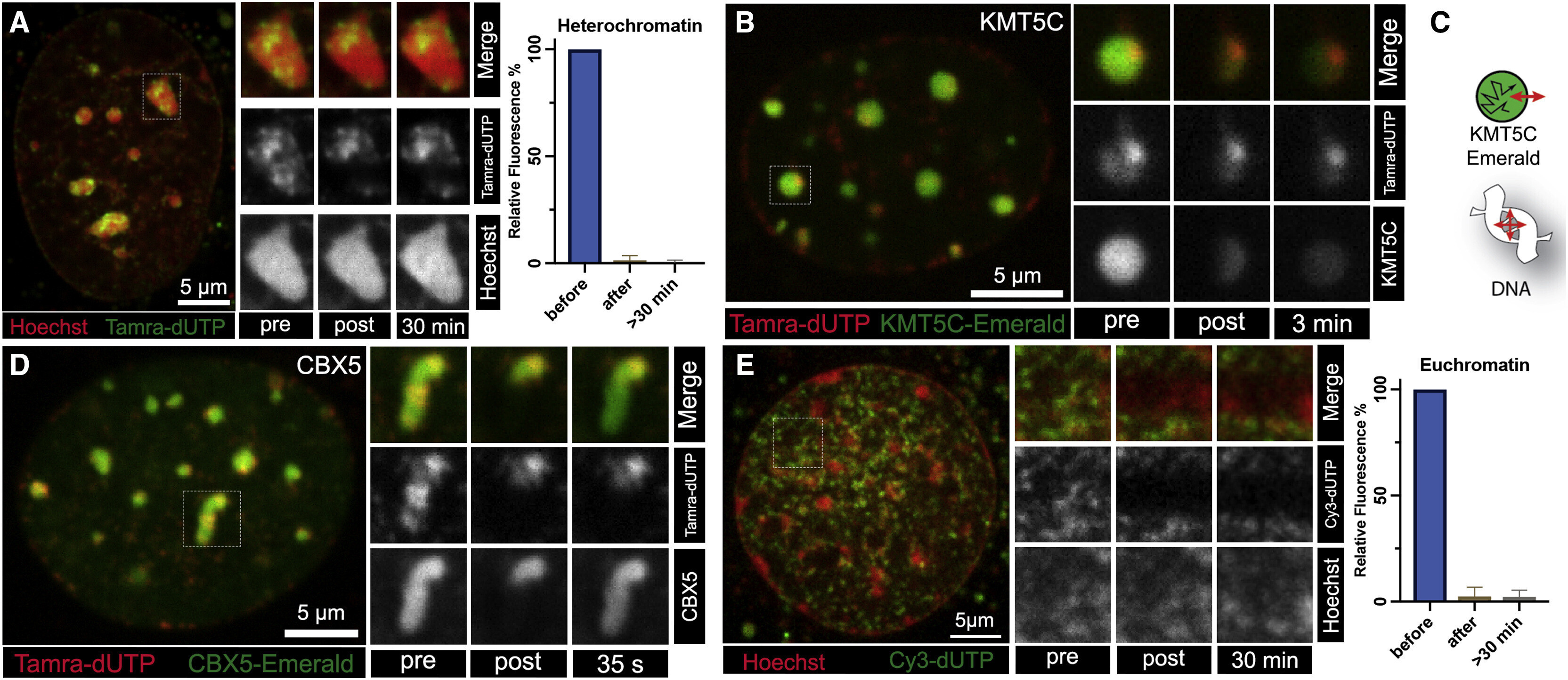
Condensed Chromatin Behaves like a Solid on the Mesoscale In Vitro and in Living Cells
Strickfaden et al.
This study examined condensed chromatin in vitro and in living cells and found solid-like behavior at the mesoscale. The results support an elastic chromatin scaffold that can coexist with liquid-like chromatin-binding proteins.
Research Imagery
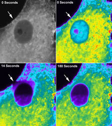
Actin Filament Structure
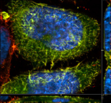
GFP-Labeled Cellular Components
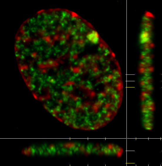
3D Nuclear Structure Projection