|
|
|
| Equipment | LSM710 | Multiphoton STED | Spinning Disc Confocal | DIMs | PALM | FCS | FLIM | TEM | Micro-Injection | HCS |Workstataions and Tools |
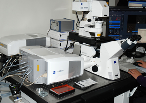
Laser Scanning Confocal Microscope (LSCM): Zeiss LSM710
The LSCM uses a point light source (laser) and a pinhole to reject out of focus light. It enables optical sectioning of fluorescently stained samples to provide a 3-Dimensional view of the structure. For more information on confocal microscopy, please go to resource section.
Main applications of the microscope:
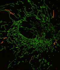
- To examine fluorescently labeled specimens for single or multiple colors localization of molecules within cell/tissue.
- To carry out physiological studies such as ratio imaging, time-lapse recording of live cell/tissue.
- Photo manipulation of the fluorescence signal (e.g. FRAP (photo bleaching after fluorescence recovering); FLIP (Fluorescence Loss Imaging after photobleaching), photoactivation/switching, etc.) to study the kinetics of molecules in living cells.
|
 |
This system is mainly used for detection of fluorescence labeling in fixed samples. However, it is also equipped with necessary accessories (temperature, CO2 controls, O2) for live cell imaging. Additionally, the system is equipped with an FCS module which also allows imaging using the sensitive Avalance Photodiode Detector (APD, Zeiss Confocor 3, see FCS section).
The instrument is equipped with a spectral detector which uses a PMT (Photon Multiplier Tube) array and optical grating elements to collect spectral information for fluorophores. Spectral information from 405nm to 720nm with step size of 10.7nm can be detected with the setup. Then, using the built-in, non-linear un-mixing tool, signals from different fluorophores can be separated mathematically. The system eliminates the need for emission filter and offers the advantage of being able to separate fluorophores with extensive emission spectra overlap. For example, GFP and YFP can be separated using this setup (which is impossible with conventional filter based imaging system). The system is also particularly useful for imaging highly auto-fluorescence sample with fluorescence imaging as auto-fluorescence has very different spectral signature than fluorophores. |
LSM 710 info:
Microscope: The Zeiss AxioObserver (inverted)
Objectives:
10x 0.45NA EC Plan-Neofluar
20x 0.8NA Plan-Apochromat
40x 1.3 oil plan-Apochromat
40x 1.2NA Water C-Apochromat
63x 1.4NA Oil DIC Plan-Apochromat
Laser lines
Diode 405nm
Diode 440nm
Argon: 458, 488, 514 nm
Solid state: 561 nm
HeNe: 633 nm
Detectors
3 fluorescence PMTs, 1 transmission PMT, and 2 APDs
|
 |
|
|