Spinning disk confocal system (PerkinElmer Ultraview ERS)
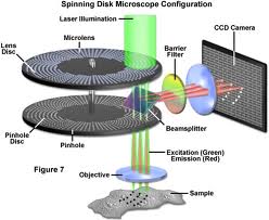
The PerkinElmer UltraVIEW system (PerkinElmer Life Sciences Inc., MA, USA) is a Yokogawa (Yokogawa Corp. Japan) Nipkow spinning disk confocal system. It uses a spinning disk with multiple pinholes to achieve confocality (e.g. the rejection of out-of-focus lights). Nipkow disk refers to scanning disk with symmetrically placed spirals of pinholes through which illumination light is passed. Such pinholes split illumination lights into multiple 'minibeams'. When the disk spins, the light scans the sample in a raster pattern. Emission lights from the sample are detected through the pinhole to generate a confocal image of the sample that can be detected (with an EMCCD (Electron Multiplification Charge-Coupled Device) camera). Because the pinholes on a Nipkow disk must be placed up to 10 diameters apart in order to avoid cross-talking problem, the light throughput of traditional Nipkow disks is only ~1% of the light shining onto the disk. The Yokogawa scanhead has overcome this problem by using an innovative, collector disk containing microlenses placed in front of the Nipkow disk. The microlenses ensure that most of the light illuminating the disk is focused onto the pinholes. Transmission efficiency is thus increased from ~1% to 70% of the light falling on the disks allowing the sample to be illuminated with a sufficient quantity of light.
|
 |
Advantages of the UltraVIEW system: The system has mainly 2 advantages over a conventional Laser Scanning confocal microscope: higher image speed and lower photo-toxicity. The Laser Scanning Confocal Microscopy (LSCM) is a sequential scanning system where a single point of the specimen is illuminated at a time and LSCM uses a point detector (Photon Multiplier Tube (PMT)). Therefore, for LSCM, an expensive scanner is needed and electronic processing is necessary for image formation. The LSCM system is consequently relatively slow (typically 0.5-10sec/frame, slower scan can take up to a minute/frame). Whereas Nipkow disk system uses multi-point scanning disk and a cooled CCD camera which is a parallel array detector. This enables the Nipkow system to acquire images at higher speed (up to 360frames/sec) than with LSCM (typically 0.5-1 frame/sec). This will allow many rapid cellular processes (e.g. calcium imaging, vesicular trafficking) to be monitored in real time. More importantly, it has been demonstrated that the spinning disk confocal system reduces phototoxicity and photobleaching by as much as 5 folds comparing to a LSCM. It is speculated that the reduction in phototoxicity and photobleaching is probably due to the fact that the system splits the laser light into thousands of minibeams.
While the mechanism of such reduction in phototoxicity and photobleaching is still a subject of studying, spinning disk confocal is becoming increasingly the instrument of choice for live cell imaging. The system is particularly powerful for applications such as real-time (4-D) confocal microscopy, calcium signaling, vesicular trafficking; green fluorescence protein studies.
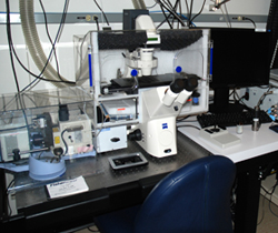
System description: The system is based on a Zeiss Axiovert 200M inverted microscope with the following laser lines for excitation of fluorophores: 404nm, 440nm, 488, 514nm, 561nm and 633nm. It is equipped with a piezo focusing motor and all necessary accessories (Temperature, CO2) for high speed live cell imaging. The scope is mounted with a high precision motorized stage for monitoring of multiple positions in a single sample holder. This system was upgraded with a FRAP (Fluorescence Recovery After Photobleaching) module in 2009.
System applications: The system is well suited for high-speed, live cell imaging with reduced phototoxicity. The system is particularly useful for 3-D timelapse to monitor fast cellular events because of the high focusing/acquisition speed than other systems in the Facility. With its FRAP module, FLIP, FRAP and photoactivation could be performed on the system as well.
|
Spinning disk confocal info:
Microscope: Zeiss Axiovert 200M (inverted) )
Objectives:
10x 0.3NA EC Plan-Neofluar
20x 0.8NA Plan Fluar
40x 1.3 oil plan-Neofluar
63x 1.2NA Water C-Apochromat
63x 1.4NA Oil DIC Plan-Apochromat
100X 1.4NA, oil DIC plan-Apochromat
Laser lines
Diode: 405nm
Diode: 440nm
Argon: 458, 488, 514 nm
Solid state: 561nm
Solid state 633nm
Detectors
Hamamatsu EMCCD 1Kx1K 8um pixel
|
 |