Cell Imaging Facility 

Department of Oncology    
Cross Cancer Institute      
Welcome
Equipment
Technologies
Using the facility
Booking
Documents
Image Gallery
Publications
Links
Contacts
Location
Equipment | LSM710 | Multiphoton STED | Spinning Disc Confocal | DIMs | PALM | FCS | FLIM | TEM | Micro-Injection | HCS | Workstataions and Tools

Fluorescence Lifetime Imaging (FLIM) module

Fluorescence is a cyclical process where lifetime of the fluorophore is not only the property of molecule itself but also a function of its environment. In situation such as Fluorescence Resonance Energy Transferring (FRET), the lifetime of donor molecule will be shortened. Therefore, FLIM can be used as a way to measure molecular interactions. Unlike conventional FRET analysis, FLIM measurement is not subjected to fluorescence photobleaching or concentration variations which are difficult to control in live cells.

The FLIM module is an attachment to the Zeiss NLO 510 system which uses time-correlated single photon counting technique to construct the decay curves of fluorophore in every pixel of the image. The system is equipped with a Hamamatsu RS-39 Multi-channel plate detector, a filter wheel and a SPC730 photon-counting board from Becker Hickl (www.becker-hickl.de) for photon counting. The system uses a photodiode to obtain synchronization information from the laser pulses to construct the fluorescence decay curve.


FLIM-Frequency_domain

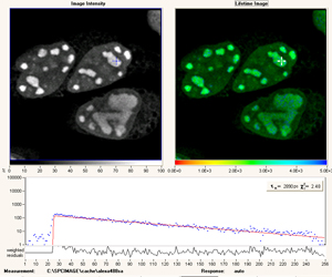
The main application for the setup in biology is for FRET analysis where donor life time can be measured both in presence and absence of acceptor molecules. Then the FRET transfer efficiency can be calculated according to the following formula:
-Et =1- t D,A/ tD

Where t D,A is the life time of donor molecule in presence of acceptor and t D is the life time of the donor molecules in absence of acceptor.

The system is integrated part of multi-photon microscope. There is no special need for specimen preparation other than that the specimen must be suitable for confocal observation. As all FRET analysis, all control samples are needed (e.g. donor alone, acceptor alone, donor acceptor combined and specimen without any staining for auto-fluorescence).
The system does not really have a user friendly interface. It is only available by assisted use. Please contact staff for more details.

FLIM


Welcome | Equipment| Technologies|Using the facility| Booking | Documents |Image Gallery|Publications|Links | Contacts|Location

Copyright 2023 Cell Imaging Facility - Department of Oncology - University of Alberta